Product Details:
|
Specimen: | Serum | Storage: | 2-8℃ |
---|---|---|---|
EXP: | 24 Months | Size: | 96 Test/Kit |
Highlight: | Anti Thyroglobulin Antibody Test,Anti Tg Ab Test Kit,BIOVANTION Ab Test Kit |
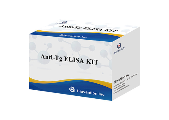
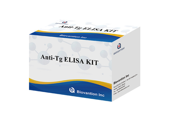
Anti-Tg ELISA TEST KIT Thyroglobulin Ab
1. Intended use
Immunoassay for the in vitro quantitative determination of antibodies to thyroglobulin in human serum. The anti‐Tg determination is used as an aid in the detection of autoimmune thyroid diseases.
2. Summary
Thyroglobulin (Tg) is produced in the thyroid gland and is a main component in the lumen of the thyroid follicle. In synergy with the enzyme thyroid‐specific peroxidase (TPO), Tg has an essential function in the iodination of L‐tyrosine and in the formation of the thyroid hormones T4 and T3. Both Tg and TPO are potentially autoantigenic.
Elevated serum concentrations of antibodies against Tg (Tg‐autoantibodies) are found in subjects with autoimmunity‐based thyroiditis. High concentrations of anti‐Tg together with anti‐TPO are indicative of chronic lymphocytic‐infiltrative thyroiditis (Hashimoto's disease). The frequency of thyroglobulin antibodies is approximately 70‐80 % in subjects with autoimmune‐thyroiditis, including Hashimoto's disease, and approximately 30 % in individuals with Graves' disease.The anti‐Tg assay is important for use in monitoring the course of Hashimoto's thyroiditis and for the differential diagnosis (cases of suspected autoimmune thyroiditis of unknown origin with negative anti‐TPO test results, Graves' disease without lymphocytic infiltration, and to rule out interference by Tg‐autoantibodies in the Tg test).
Although the sensitivity of the procedure can be increased by simultaneously determining additional thyroid antibodies (anti‐TPO, TSH‐receptor‐antibodies), a negative result does not definitively rule out the presence of an autoimmune disease. The level of the antibody titer does not correlate with the clinical activity of the disease. Titers that are elevated initially can become negative if the disease persists for a longer period of time or if
remission occurs. If antibodies reappear after remission, a relapse is likely.
The Enzyme linked immunosorbent assays uses human antigen and rabbit antihuman IgG antibodies (anti-IgG).
3.Reagents Materials provided
Coated Microplate, 8 x 12 strips, 96 wells. Pre-coated with human TG antigen.
Calibrators, 6 vials, 1 mL each, ready to use; Concentrations: 0(A), 50(B) ,150(C), 500(D), 1000(E) and 2000(F) IU/mL.
Enzyme Conjugate, 1 vial, 11.0 mL of HRP (horseradish peroxidase) labeled rabbit anti- human IgG antibodies (anti-IgG) in Tris-NaCl buffer containing BSA (bovine serum albumin). Contains 0.1% ProClin300 preservative.
Serum Diluent: 1 vial, 11mL. Containing buffer salts and a dye
Wash Solution Concentrate, 1 vial, 25 ml (40X concentrated), PBS-Tween wash solution.
Substrate, 1 vial, 11ml, ready to use, (tetramethylbenzidine) TMB.
Stop Solution, 1 vial, 6.0 ml of 1 mol/l sulfuric acid.
IFU, 1 copy.
Plate Lid: 2 pieces.
4. Materials required (but not provided)
5. Test procedure
Use only the number of wells required and format the microplate wells for each calibrator and sample to be assayed.
Add 100 µL of calibrators to each well.
Add 100 µL of Sample Diluent (Green color) to each well Except the Calibrator-wells.
Add 10 µL of Sample to each Sample Diluent well (NOTE: Reagents in Wells will turn Blue color from Green), then shake 30 seconds.
Cover the plate with a plate lid and incubate at 37 °C for 30 minutes.
Discard the contents of the micro plate by decantation or aspiration. If decanting, tap and blot the plate dry with absorbent paper.
Add 350 µL of wash solution, decant (tap and blot) or aspirate. Repeat 4 additional times for a total of 5 washes. At the end of washing, invert the plate and tap out any residual wash solution onto absorbent paper.
Add 100 µL of enzyme conjugate,
Cover the plate with a plate lid and incubate at 37 °C for 30 minutes
Add 350 µL of wash solution, decant (tap and blot) or aspirate. Repeat 4 additional times for a total of 5 washes. At the end of washing, invert the plate and tap out any residual wash solution onto absorbent paper.
Add 100 µL of Substrate to each well. Cover and incubate at ambient temperature (18-25℃) in the dark for reaction for 10 minutes. Do not shake the plate after substate addition.
Add 50 µL of stop solution to each well.
Shake for 15-20 seconds to mix the liquid within the wells. It is important to ensure that the blue color changes to yellow completely.
Read the absorbance of each well at 450 nm ( using 620 to 630 nm as the reference wavelength to minimize well imperfections) in a micro plate reader. The results should be read within 30 minutes of adding the stop solution.
Contact Person: Mr. Steven
Tel: +8618600464506